Biology 297C
Gel Permeation Chromatography
by Joe Kunkel
Introduction
The purpose of this experiment is to provide an introduction
to the exceedingly useful subject of column chromatography. Only one of the
many variations of this method of fractionation of different molecules will
be used here, but, in practice, a combination of several types is often
the preferred approach for separating macromolecules.
We will investigate the use of gel permeation chromatography.
One note on terminology, this same technique is often referred to as gel
filtration, a misnomer. The descriptive, filtration, refers to a technique used
to divide a sample into two fractions. The same media used for gel permeation
chromatography can be used for gel filtration. When filtration is the objective
the desired product is usually designed to come off the column first in the void
volume, Vo, or excluded volume, that volume that resides outside
the gel. While this is certainly a powerful use of the media, it is in no way
chromatography which implies spreading the desired product across a spectrum
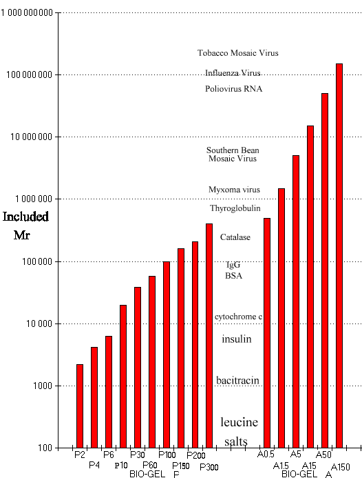
of differences. To see the spectrum of molecules separable by gel permeation
chromatography examine the distribution of some molecules separated by the
BioRad P and A series of gel beads, Fig 1.
Procedure
Each group has been provided with the raw materials to build a gel column: a glass column, BioRad P-x (where x = 100, 200 or 300) gel to fill the column, various connectors, a ring stand, clamps and test tubes to collect your product, and an appropriate amount of gel beads. You will build your column on day one of the exercise, then store it until the following lab when it will be packed with gel and used to separate a supplied mixture of molecules.
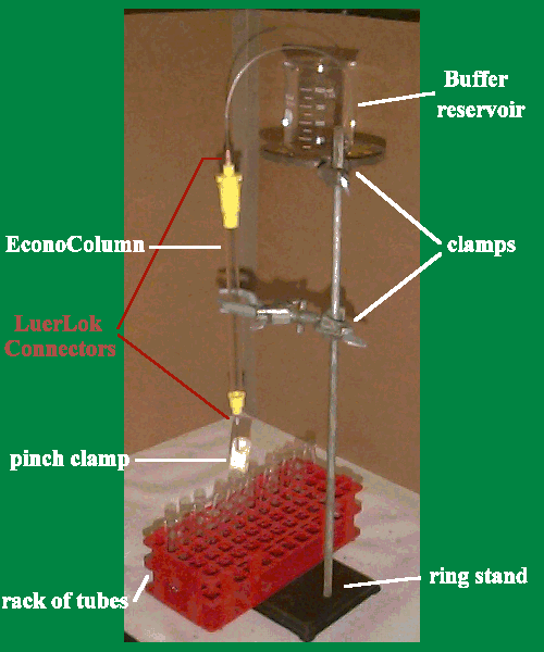
Place a Luer-Lok blunted 18 gauge needle into the bottom of the provided EconoColumn and attach a length of narrow tubing with a pinch clamp to the tip; this will allow you to stop the flow of elluent that exits the bottom of your column. The gel column will rest on the built-in fritted disc visible at the base of the glass walls of your column. This fritted disc ensures that the fine gel permeation beads do not leak out of the bottom of the column. It is desirable to have the bottom and top of the gel column surface level. Fig 2 illustrates the assembled column.
Assemble the column securely in a vertical position on
the ring stand; allow room for the test tubes for collecting fractions
below the column outlet. (It is advisable to assemble the collection tubes
sequentially in a test tube rack and to move the entire rack from fraction
to fraction beneath the column outlet tube).
Obtain a small amount of the BioRad P-x slurry. (10-30
ml. depending on the bed volume of your column). Try to agitate this as
little as possible in order to avoid introducing air bubbles into the gel.
Obtain about 200 ml of the phosphate buffered saline which will be used
for elution of the column. Mark the column height every 2 cm. with a glass
marking pencil starting from the bottom of the column bed. Close the outlet.
Pour buffer into the column to a height of about 2 cm. It is desirable
to have a column about 25 cm. high and it is also advisable to do all of
the pouring of the slurry before the gel settles. Settling of the gel between
additions results in banding of the gel which creates turbulence in the
flow. Fill the column with slurry to a height of 20-25 cm preferably by
pouring down a glass rod. If any air bubbles have been introduced carefully
attempt to remove them by tapping the column gently or by dislodging them
with a thin glass stir rod. Open the outlet briefly in order to make certain
that the effluent buffer is clear and contains no gel particles.
Ideally, a BioRad P-x bed would be allowed to settle for
several hours (under slightly reduced pressure if the column were long);
however, Time may not permit this treatment here. Instead, gently apply
a small reservoir of buffer above the slurry, replace the stopper securely
in the top of the column, and attach tubing from the elevated buffer reservoir
to the top opening. (It may be necessary to fill the tubing by gently applying
suction to start the siphon action). Open the column outlet and wash the
column, at the same time permitting it to settle. Collect the effluent
in a clean beaker and return it to the reservoir. Control the rate of column
flow by the height of the upper reservoir; do not lower the reservoir below
the column outlet as flow will be reversed. The column should flow at about
1 or 2 drops per second. A faster flow rate is undesirable and will pack
the column too tightly. If the column settles below 20-25 cm., it is necessary
to add move slurry. This is done by disconnecting the reservoir, gently
stirring the top of the column bed and adding slurry. If the column bed
is too long, the gel can be removed with a disposable pipette (replace
this gel into your supply). After doing any adjustments to your column
bed, place a few cm. of buffer on top of the column and gently stir in
order to have a level column top. After any adjustments, again connect
the buffer reservoir and wash the column for 5-10 minutes.
While the column is washing prior to chromatography, calibrate 20 test tubes to contain 1 ml. each. Do this by adding 1 ml. of distilled water, marking the level, and discarding the water. Number the tubes and use them to collect the chromatographic fractions.
When the column bed height no longer changes, settling
is complete. At this time prepare to apply the sample which is to be
chromatographed.
It should be stressed that at all times, care must be taken to ensure that
the level of buffer never falls below the level of the top of the column
bed.
Obtain 0.5 ml. of the sample mixture which is to be separated.
In another clean beaker place 5 to 10 ml. of the buffer from the reservoir;
this will be used for washing the sample onto the column and for replacing
the reservoir on top of the bed. A separate source of the same buffer is
used during these operations to ensure that the large reservoir does not
become contaminated by the chromatographic sample during these operations.
Prepare the column for sample addition by disconnecting
the reserevoir and removing the small reservoir on top of the column bed.
This last manipulation is best done by piperting off most of the buffer
with a disposable pipette; the remainder is removed by opening the outlet
and carefully drawing off buffer to the level of the top of the column
bed. Using a disposable pipette carefully apply the sample symmetrically
on top of the bed. This is best done by adding it dropwise around the
circumference
of the bed while bracing the pipette against the side of the glass column.
The object is to place the sample on the column bed and not in it, so do
not forcefully expel sample from the pipette. Open the outlet and let the
sample run onto the column flush to the top of the gel. With the outlet
closed, "wash" the sample on by adding, as carefully as you did
with sample, 0.5 ml. of column buffer. Apply this in exactly the same fashion.
After this is applied, close the bottom stopcock, very gently layer several
mls. of buffer on top of the column, connect the reservoir, open the outlet
and begin serial collection of 1 ml samples. After the sample is applied,
the longer that the column is not running, the more diffusion (spreading)
of the sample band there will be. For this reason work quickly, but not
at the cost of care and precision.
While the column is running try to observe the separation of the sample into bands; remember to change the collection tube after each 1 ml. of effluent. Alter a full columns volume (+1 ml) has come off of the column close the stopcock and disconnect the reservoir. Take the column down and empty the BioRad Px back into your supply beaker. The column can be easily emptied by blowing gently on the outlet tube while inverting it over the beaker. Return the gel to the instructor to replace in the supply.
Treatment of Results
Note your sample collection tubes, and using a scale of
0 to 4, visually estimate the color and intensity in each of the tubes by
placing a white card behind the tube. Record
these values in a table. Prepare a chromatogram by plotting tube number
on the abscissa and color 'intensity on the ordinate. On this plot be sure
to specify the chromatography conditions including: column size, sample
size, fraction size, eluting buffer, and hydrostatic pressure (height from
top of reservoir to the bottom of the column outlet). You will further use the
Bradford Protein assay and UV absorbance to quantify the amount of protein
in the 1 ml fractions
you have collected. Add the results to the table of visible products.
Discussion
(1) What is the void volume, V0, of your BioRad P-x column? This gel has an exclusion limit molecular weight of about ______.
(2) Describe the appearance of the sample bands as they separated on the column. If there was any skewing of the bands, attempt to explain this. What factors during elution can lead to imperfections in chromatographic separations on BioGel Px?
(3) Hemoglobin has an isoelectric point of 6.9 while that of cytochrome c is 10.6. On the basis of this, in what order would you predict that they would be eluted from Amberlite IRC - 50 (a polycarboxylic acid resin) using phosphate buffer at pH 6.9 as an eluent? What are the reasons for your prediction?
APPENDIX
GEL PERMEATION CHROMATOGRAPHY HISTORY
An early observation of the molecular sieve effect in
gels was made by Synge and Tiselius who fractionated hydrolysis products
of amylose in agar-agar jelly. More recently, fractionation of biological
materials has been accomplished on Sephadex, granulated agar, polyacryamide,
pelletized cellulose, cross-linked hyaluronic acid, polyvinyl ethyl carbitol,
and polyvinyl pyrolidone.
Porath and Flodin introduced particular dextran gels for
desalting protein in 1959. Dextran is a heterogeneous glucan of high molecular
weight produced by many strains of Leuconostoc. Most of the glucosidic
linkages are alpha-D-1->6 but a few are of the 1->3 type. A stable
three dimensional polymer can be formed from dextran by reaction of the
glucose hydroxyls with the cross-linking agent epichlorohydrin or with
bifunctional epoxides. Two alcohol molecules are bridged in the following
manner with little change in the total hydroxyl content
R - OH + CH2 - CH - CH2
- C1 + H0 - R
\_O_/
R - O - CH2 - CH (OH) CH2 - O - R'
The cross-linking results in a three dimensional network
of polysaccharide chains which, in a simple view, functions as a sieve.
Molecules larger than the largest pores of the beads cannot penetrate the
gel particles; and thereafter they pass through the bed in the liquid phase
outside of the particles. They are not impeded and are eluted first. Smaller
molecules penetrate the gel particles to a varying extend depending on
their size and shape. Molecules are therefore, eluted from a gel permeation
bed in the order of decreasing molecular size. As the molecules pass through
the bed at different rates, they emerge at the outlet of the column separated
from each other. The zones eluted are somewhat wider than the sample applied.
The basic equation describing the operational parameters
is
Ve = Vo + Kd Vi
where Ve is the elution volume, Vo is the void volume, Vi the volume of the water imbibed by the gel particles, and Kd the distribution coefficient. Kd bears a resemblance to the similar expression used in normal chromatography, the partition coefficient, which is the ratio between the concentration of a solute in the mobile and stationary phases. In gel filtration the stationary phase may be considered to be the water imbibed by the gel particles and Kd the fraction of the imbibed water available, for distribution of the solvent. The actual concentration of the solute interior to the gel pores may be assumed to be practically identical with that of the external liquid phase. Knowing the weight of dry gel a, and the water regain Wr. Kd can be determined from experimental data using the relation
              Ve - Vo      Ve - Vo
      Kd =    -------  =  --------
                 Vi         a Wr.
This brief outline of a single technique must not exclude
an awareness of the several other equally valuable methods of chromatographic
separation of substances. An equally, if not more, important method in
this area is ion exchange chromatography. It is essential to consult the
cited literature and the text in order to be aware of these additional
techniques.